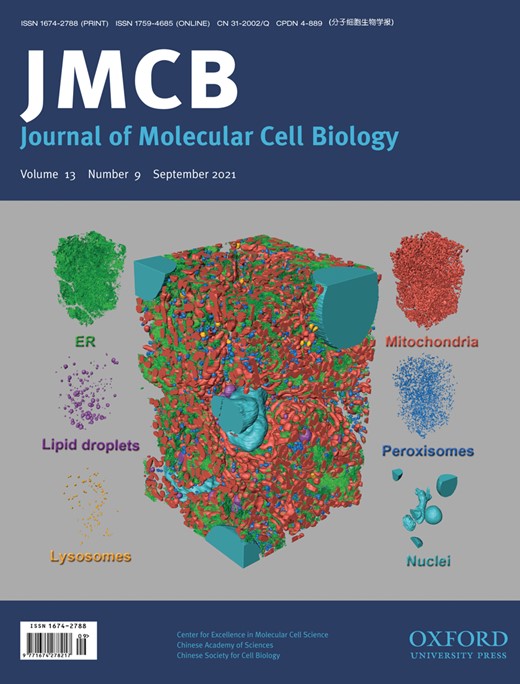
The endoplasmic reticulum (ER) is a contiguous and complicated membrane network in eukaryotic cells, and membrane contact sites (MCSs) between the ER and other organelles perform vital cellular functions, including lipid homeostasis, metabolite exchange, calcium level regulation, and organelle division. Here, we establish a whole pipeline to reconstruct all ER, mitochondria, lipid droplets, lysosomes, peroxisomes, and nuclei by automated tape-collecting ultramicrotome scanning electron microscopy and deep learning techniques, which generates an unprecedented 3D model for mapping liver samples. Furthermore, the morphology of various organelles and the MCSs between the ER and other organelles are systematically analyzed. We found that the ER presents with predominantly flat cisternae and is knitted tightly all throughout the intracellular space and around other organelles. In addition, the ER has a smaller volume-to-membrane surface area ratio than other organelles, which suggests that the ER could be more suited for functions that require a large membrane surface area. Our data also indicate that ER‒mitochondria contacts are particularly abundant, especially for branched mitochondria. Our study provides 3D reconstructions of various organelles in liver samples together with important fundamental information for biochemical and functional studies in the liver.